What Is Cervical Fusion Surgery?

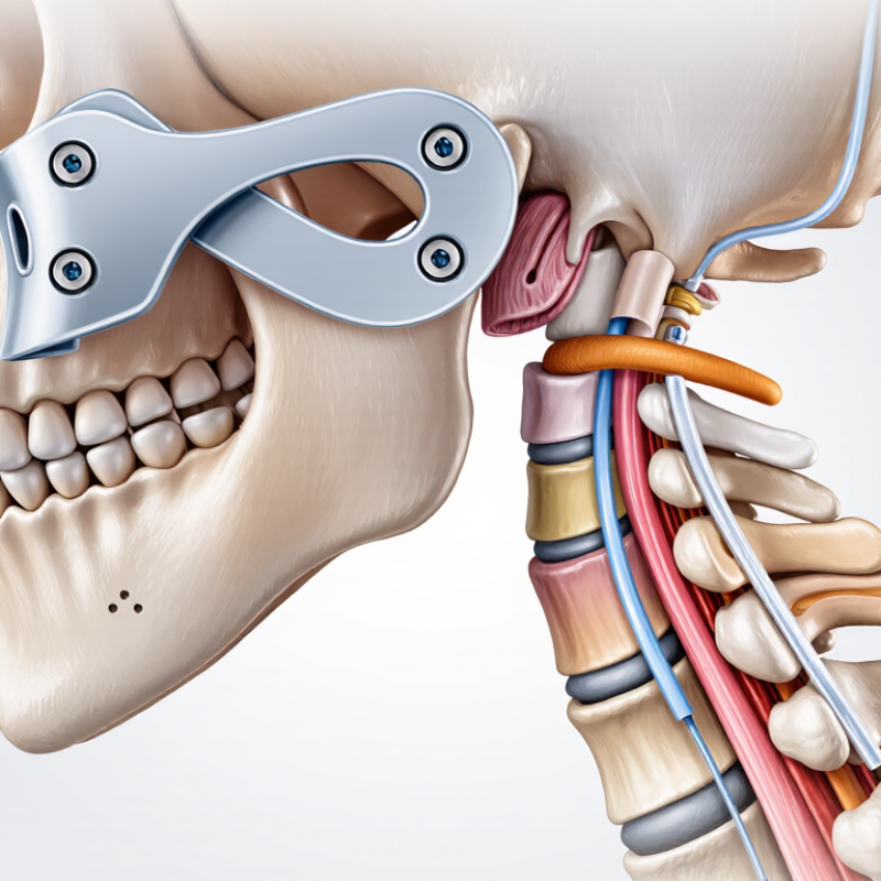
Cervical fusion surgery is a procedure that joins two or more neck vertebrae. The goal is to reduce painful movement and protect irritated nerves or the spinal cord. A surgeon may remove a damaged disc, place a bone graft or implant, and stabilize the area with plates or screws. This process is commonly called cervical fusion.

People may consider this operation after persistent neck pain, arm weakness, numbness, or balance problems. These symptoms can result from a herniated disc, spinal stenosis, arthritis, or injury. However, symptoms alone do not determine whether surgery is appropriate. A careful evaluation usually includes a medical history, physical examination, and imaging such as an MRI or X-ray.

The decision deserves time.

Cervical fusion can improve nerve-related symptoms, but it is not a guaranteed cure for every type of neck pain. Recovery may involve temporary stiffness, swallowing discomfort, activity limits, and physical therapy. Rare but serious complications include infection, nerve injury, blood clots, or problems with the fusion healing properly. Long-term stress may also affect nearby spinal levels, although this does not happen to everyone.

An experienced spine specialist should explain the expected benefits, alternatives, and personal risks. Patients should also discuss smoking, bone health, medications, and previous surgeries. These details can influence healing. Medical advice varies because each neck is different. Even standard treatment plans may need adjustment. This guide explains what cervical fusion involves, why it may be recommended, and what patients should realistically expect before, during, and after surgery.

What Is Cervical Fusion Surgery?

What Is Cervical Fusion Surgery? Anatomy of the Seven Cervical Vertebrae

Cervical fusion surgery joins two or more neck vertebrae to limit painful movement. The cervical spine contains seven vertebrae, labeled C1 through C7. C1 is the atlas, which supports the skull. C2 is the axis, allowing much of the head’s rotation. C3 through C7 share a more typical structure. Each has a vertebral body, an arch, and an opening for the spinal cord. Small openings also allow nerves and blood vessels to pass. Most levels contain flexible discs, but C1 and C2 do not.

These bones form a strong yet mobile column. Facet joints guide movement between neighboring vertebrae. The spinal cord travels through the central canal, while nerve roots exit through side openings. Narrowing near C5-C6 or C6-C7 may irritate nerves reaching the shoulder, arm, or hand. Symptoms can include pain, tingling, weakness, or reduced reflexes. Anatomy is not perfectly symmetrical. That matters.

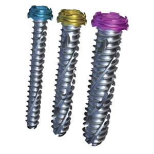
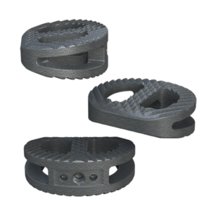
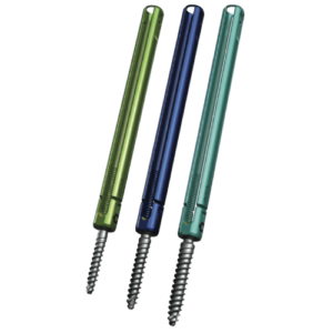
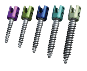
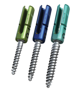
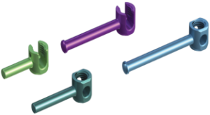
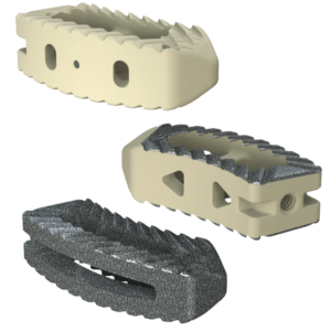
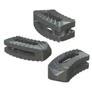
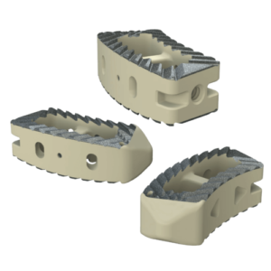
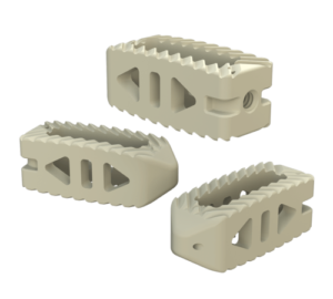
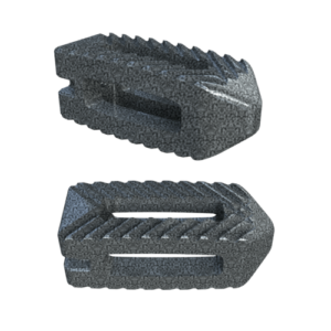
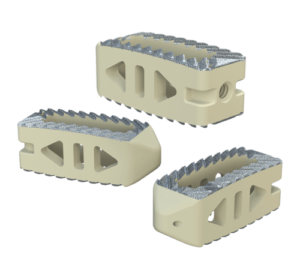
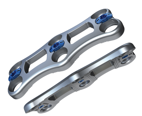
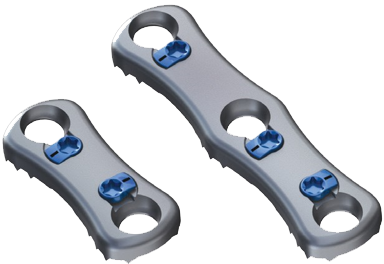
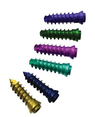
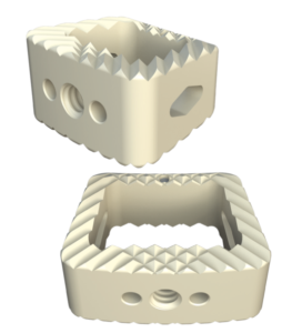
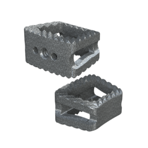
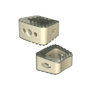
During fusion, a surgeon removes damaged disc material or bone pressing on neural tissue. A spacer or bone graft then helps create a solid connection between selected vertebrae. Plates, screws, or rods may provide temporary stability while healing occurs. The procedure can reduce motion at one level, but it does not repair every source of neck pain. Careful examination and imaging are essential because similar symptoms may arise from muscles, nerves, or the spinal cord. Recovery varies, and some uncertainty remains even after technically successful surgery.

Why Is It Recommended? Degeneration, Trauma, Instability, and Nerve Compression

What Is Cervical Fusion Surgery?

Cervical fusion surgery joins two or more neck vertebrae. Surgeons remove a damaged disc or bone, then place a spacer and fixation to stabilize the spine. The procedure may be recommended when pain, weakness, or spinal cord symptoms persist despite appropriate non-surgical care.

Degeneration is common. A 2015 review in the American Journal of Neuroradiology found disc degeneration on scans in 37% of adults aged 20 and 96% of adults aged 80, including many people without symptoms. An abnormal scan alone is not enough.

Clinical reasoning matters.

Trauma can fracture cervical bones or damage supporting ligaments. The World Health Organization reports 20–50 million nonfatal road-traffic injuries worldwide each year. Some injuries create instability, making ordinary movement unsafe. Fusion may restore structural support when bracing or other treatment cannot control the risk.

Nerve compression from a herniated disc, bone overgrowth, or narrowed foramina can cause arm pain, numbness, or weakness.

Spinal cord compression may produce clumsy hands, balance problems, or stiff legs.

Not every painful neck needs fusion. The North American Spine Society emphasizes matching imaging findings with symptoms and examination results. This step is often underestimated.

Surgery also carries risks, including infection, swallowing difficulty, adjacent-level degeneration, and nonunion. Patient age, smoking, bone health, neurological changes, and injury severity can alter the decision. A second clinical opinion may be sensible when symptoms and scans do not align.

How Is ACDF Performed? Steps and the 85–95% Fusion-Rate Range

What Is Cervical Fusion Surgery?

Anterior cervical discectomy and fusion, or ACDF, treats pressure on cervical nerves or the spinal cord. Surgeons usually recommend it after imaging, neurological examination, and failed conservative care. The operation begins under general anesthesia. The patient lies face-up, with the neck gently extended.

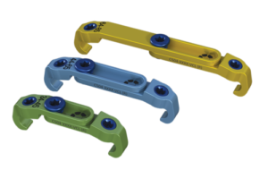
Through a small front-of-neck incision, the surgeon moves the windpipe and esophagus aside. The damaged disc and nearby bone spurs are removed. This creates space for the compressed nerve or spinal cord. A bone graft or structural implant then fills the disc space. Plates and screws may provide immediate stability. X-ray imaging helps verify alignment before closure. The entire process often takes one to several hours, depending on the number of levels.

The range matters. Peer-reviewed reviews in Spine and the Journal of Neurosurgery: Spine commonly report fusion rates of approximately 85–95% after ACDF, especially in single-level procedures. Rates can decline with multilevel surgery, smoking, diabetes, poor bone quality, or inadequate follow-up. Professional spine literature also warns that radiographic fusion does not always equal complete symptom relief. That distinction deserves attention. A healed fusion may still leave numbness or weakness if nerve damage was longstanding. Conversely, some patients improve before solid fusion appears on imaging. Follow-up scans, often taken months later, assess bone bridging and hardware position. The 85–95% figure is useful, but it is not a promise.

What Is Cervical Fusion Surgery?

How Is ACDF Performed? Steps and the 85–95% Fusion-Rate Range

Anterior cervical discectomy and fusion (ACDF) is a cervical spine procedure that removes a damaged disc from the front of the neck, relieves pressure on the spinal cord or nerve roots, and stabilizes the treated segment with a bone graft or spacer.

  1. A small incision is made at the front of the neck.
  2. The surgeon gently reaches the cervical spine and identifies the affected disc.
  3. The damaged disc and any compressive bone or tissue are removed.
  4. A bone graft or interbody spacer is placed in the disc space.
  5. Additional stabilization may be provided with a plate and screws.
  6. The incision is closed while the graft gradually fuses the adjacent vertebrae.

The chart shows the commonly cited 85–95% fusion-rate range. Actual results vary according to the number of levels treated, patient health, bone quality, surgical factors, and follow-up duration; these values are not a guarantee for an individual patient.

Which Techniques Are Used? Anterior, Posterior, and Multilevel Fusion

What Is Cervical Fusion Surgery?

Which Techniques Are Used? Anterior, Posterior, and Multilevel Fusion

Cervical fusion surgery joins two or more neck vertebrae to reduce painful movement and stabilize the spine. Surgeons may remove a damaged disc, bone spurs, or other pressure sources around the spinal cord and nerves. A bone graft and fixation device then help the vertebrae grow together. Fusion is not simply a way to “repair” the neck. It permanently limits movement at the treated level.

The anterior approach reaches the spine through the front of the neck. It commonly allows direct removal of a damaged disc or bone growth. Many patients experience less muscle disruption with this route, but swallowing discomfort can occur temporarily. The posterior approach enters through the back of the neck. It may suit cases involving spinal narrowing, instability, or several affected levels. Muscle soreness can be more noticeable after surgery.

Multilevel fusion treats two or more cervical levels during one operation. It can provide stronger overall stabilization, yet it may require a longer recovery and create greater stress on nearby segments. No technique fits every patient. Not even close. A surgeon reviews symptoms, neurological findings, imaging, bone health, and previous operations before choosing an approach. Recovery varies, and pain relief is not guaranteed. Rare but important complications include infection, nerve injury, nonunion, and breathing or swallowing problems. I would not treat a scan alone; the patient’s daily function matters just as much.

What Do Outcomes Show? Recovery Timelines and Reported Complication Rates

Cervical fusion surgery removes a damaged disc or stabilizes painful neck segments with bone graft material. Outcomes depend on diagnosis, spinal levels treated, age, smoking, and general health. Clinical studies commonly report meaningful relief of arm pain in about 70% to 90% of patients. Neck pain may improve more slowly and less predictably. Recovery is not linear. Some patients walk the same day, while others need several days of supervised care.

Many people resume desk work within two to six weeks. Heavy lifting and driving may require longer restrictions. Bone fusion usually develops over three to six months, although imaging can show delayed healing. Early swallowing discomfort affects roughly 5% to 20% of patients, especially after front-of-neck surgery. It usually improves within days or weeks. Persistent swallowing problems appear less often, commonly around 2% to 5% in published reports. Infection, nerve injury, and blood-clot complications are uncommon, often reported below 1% to 2%. Nonunion rates may range from 2% to 10%, with higher figures after multilevel surgery or nicotine exposure. These numbers are not perfectly comparable. Studies define complications differently, and follow-up periods vary. The statistics can look cleaner than real life. A careful surgeon still discusses unexpected pain, delayed healing, and possible revision surgery before treatment.

What Is Cervical Fusion Surgery? – What Do Outcomes Show? Recovery Timelines and Reported Complication Rates

Data Dimension Typical Finding or Reported Range What It Means Important Considerations
Purpose of cervical fusion Removal of a painful or compressive disc, followed by stabilization of adjacent vertebrae The procedure is intended to relieve nerve-root or spinal-cord compression and create a solid fusion between vertebrae. Common indications include cervical radiculopathy, myelopathy, instability, deformity, fracture, or degenerative disc disease that has not improved with appropriate nonsurgical treatment.
Common surgical approach Anterior cervical discectomy and fusion is frequently used for one- or more-level disease; posterior fusion may be selected for different patterns of compression or instability The approach depends on the location of compression, number of affected levels, spinal alignment, and the surgeon’s assessment. Outcomes and complication risks should not be directly compared across procedures without considering these differences.
Neck and arm pain improvement Approximately 70–95% of appropriately selected patients report meaningful improvement Arm pain caused by nerve compression often improves more predictably than nonspecific neck pain. Results vary according to diagnosis, duration of symptoms, smoking status, psychological health, worker demands, and the presence of spinal-cord damage.
Neurologic improvement in myelopathy Approximately 60–90% show improvement or stabilization Surgery commonly prevents further deterioration and may improve walking, hand function, balance, or abnormal reflexes. Long-standing spinal-cord injury may be only partly reversible. Earlier treatment is often associated with a better chance of recovery.
Radiographic fusion success About 90–99% for many single-level anterior procedures; approximately 70–95% for multilevel procedures A successful fusion means that bone has formed across the treated level or levels. Fusion rates depend on the number of levels, bone quality, surgical technique, graft material, smoking, diabetes, and medication factors.
Early postoperative pain Usually greatest during the first several days and improves substantially over 2–6 weeks Incisional neck pain, muscle tightness, shoulder discomfort, and temporary swallowing discomfort are common. New or worsening weakness, severe difficulty swallowing, breathing problems, fever, or loss of bladder or bowel control requires urgent medical evaluation.
Return to desk-based work Approximately 2–6 weeks Many patients can resume light computer or administrative work once pain is controlled and medications do not impair alertness. The timing depends on fatigue, neck mobility, commuting, work ergonomics, and whether complications occur.
Return to physically demanding work Approximately 6–12 weeks or longer Lifting, repetitive neck movement, pushing, pulling, and vibration may require a longer restriction period. Work clearance should be individualized and should follow the operating surgeon’s restrictions.
Time to biological fusion Often 3–6 months; some multilevel fusions require 6–12 months Symptoms may improve before the fusion is fully visible on imaging. Follow-up imaging is used to assess alignment, hardware position, and progression toward fusion.
Postoperative dysphagia Approximately 5–20% during the early recovery period Temporary difficulty swallowing is one of the most frequently reported complications after anterior cervical surgery. The reported rate varies with the definition used, the timing of assessment, number of surgical levels, reoperation status, and surgical technique.
Persistent dysphagia Approximately 2–5% beyond 6–12 months in many series Most early swallowing symptoms improve with time, but a smaller proportion persists. Persistent or worsening swallowing difficulty should be assessed, particularly if accompanied by coughing, aspiration, weight loss, or breathing symptoms.
Pseudarthrosis or nonunion About 1–5% for many single-level procedures; approximately 5–20% for multilevel procedures Pseudarthrosis means that the intended bony fusion has not become solid. Risk increases with more treated levels, smoking, poor bone quality, diabetes, and certain medication or nutritional factors. Some patients have no symptoms; others require additional treatment.
Adjacent-segment degeneration or disease Symptomatic adjacent-level disease has been reported at roughly 2–3% per year in long-term observational data Levels next to the fusion may later develop radiographic degeneration or new symptoms. This may reflect the natural history of cervical degeneration as well as altered biomechanics. Radiographic changes do not always cause symptoms or require surgery.
Reoperation after cervical fusion Approximately 5–15% over several years, depending on follow-up duration and procedure type Additional surgery may be needed for nonunion, recurrent or adjacent-level compression, hardware problems, infection, or persistent symptoms. Longer follow-up and multilevel surgery generally increase the observed cumulative reoperation rate.
Nerve-root or spinal-cord injury Usually well below 1% in modern elective surgical series New neurologic injury is uncommon but potentially serious. Risk depends on the underlying disease, severity of compression, surgical approach, anatomy, and emergency versus elective status.
Infection Approximately 0.5–2% Most infections are uncommon after elective cervical fusion but may require antibiotics, drainage, or further surgery. Diabetes, obesity, smoking, immune suppression, prolonged surgery, and revision procedures may increase risk.
Blood clot or pulmonary embolism Generally below 1% after elective cervical fusion Venous thromboembolism is uncommon but can be life-threatening. Risk is influenced by age, cancer, immobility, prior clotting events, obesity, and other medical conditions.
Overall interpretation of outcomes Most appropriately selected patients experience improvement in arm pain, neurologic function, or both Cervical fusion is generally effective for nerve compression and spinal-cord compression when the diagnosis and surgical indication are appropriate. Clinical improvement, radiographic fusion, and complete elimination of pain are different outcomes; a solid fusion does not guarantee that every symptom will resolve.

How to read these figures: The percentages are approximate ranges compiled from published clinical studies and systematic reviews. Reported rates differ because studies use different definitions, follow-up periods, patient populations, surgical approaches, and numbers of treated levels.

Selected evidence sources: Hilibrand AS et al., long-term study of adjacent-segment disease after cervical arthrodesis; systematic reviews and meta-analyses evaluating dysphagia after anterior cervical surgery; published cohort studies and reviews reporting fusion, pseudarthrosis, neurologic, infection, and reoperation outcomes after cervical decompression and fusion.