7 Best Bone Graft Materials for Dental Implants?

Choosing a bone graft for implant placement is not a simple search for the “strongest” material. The right option depends on defect size, bone quality, healing time, soft-tissue coverage, and the patient’s medical history. A narrow ridge may need carefully shaped particles and a membrane. A small extraction socket may require a different approach. Clinical judgment matters.

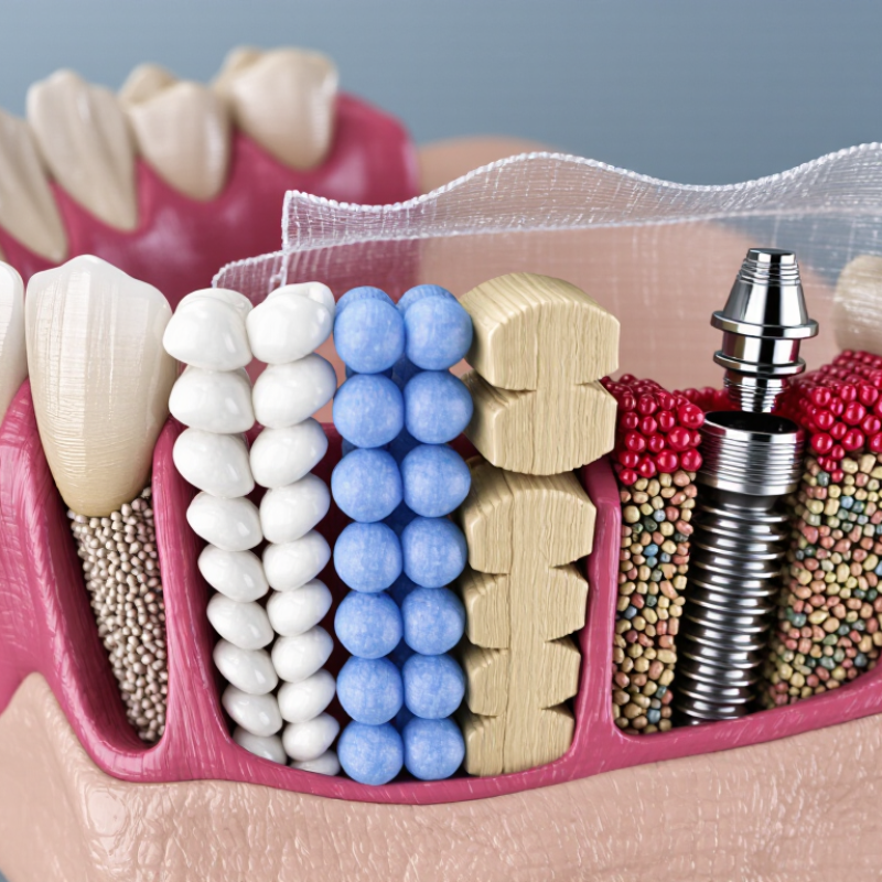
Professor Daniel Buser, a widely recognized implant dentistry expert, described the purpose of augmentation as creating “an ideal three-dimensional bone contour around the implant.” That principle keeps the focus on function, stability, and long-term tissue support. Materials commonly discussed include autogenous bone, human allografts, animal-derived xenografts, synthetic alloplasts, dentin-based grafts, platelet concentrates, and composite grafts. Each has practical advantages and limitations. Some materials resorb quickly. Others preserve volume longer. Neither outcome is automatically better.

The word “best” deserves caution. It can oversimplify a biological decision. Evidence supports several materials, but results also depend on surgical technique, infection control, healing conditions, and maintenance. This guide compares seven leading bone graft materials for implant treatment, using their sources, handling properties, remodeling behavior, and clinical applications. It also considers patient concerns, including treatment time, religious preferences, cost, and material acceptance. Small details matter, such as how firmly particles remain beneath a membrane or how the graft looks during follow-up imaging. No material guarantees success. Careful diagnosis does.

7 Best Bone Graft Materials for Dental Implants?

Clinical criteria for ranking seven graft materials: defect size, biology, and risks

7 Best Bone Graft Materials for Dental Implants?

There is no universal winner. The best graft depends on defect size, healing biology, implant timing, and patient risk. In clinical practice, autogenous bone often ranks highest for challenging defects because it provides living cells and growth signals. However, harvesting it may cause pain, bleeding, or limited volume.

For moderate defects, freeze-dried human bone can offer useful structure without a second surgical site, although disease screening and gradual remodeling remain important. Animal-derived mineral grafts maintain space well but may remodel slowly. Synthetic hydroxyapatite, beta-tricalcium phosphate, biphasic calcium phosphate, and bioactive glass provide additional options. Their handling, porosity, strength, and resorption rates differ. A collagen sponge may support small contained defects, but it is not a strong choice for large vertical losses. Size matters. Biology matters more than popularity. Smoking, uncontrolled diabetes, infection, thin soft tissue, and poor blood supply can change the ranking completely. I would not treat a material label as a treatment plan.

Tips: Match the graft to the defect, not the advertisement. Measure width, height, containment, and membrane stability with clinical examination and imaging. For a small socket, a resorbable scaffold may be enough. For a wide, non-contained defect, a stronger space-maintaining approach may be necessary. Review medical history carefully, explain realistic healing times, and reassess if early inflammation appears. Even experienced clinicians can misjudge the defect before flap elevation.

Autogenous bone: osteogenic potential and typical 4–6-month healing periods

Among the best bone graft materials for dental implants, autogenous bone remains clinically important. It comes from the patient’s own jaw, chin, or hip area. This tissue contains living bone-forming cells, giving it genuine osteogenic potential. It can also provide a natural framework for new bone growth. That combination makes autogenous grafting biologically attractive.

It is not always the easiest choice. A second surgical site may cause soreness, swelling, or temporary numbness. In practice, patients often notice discomfort near the harvest area before the implant region. Healing commonly takes about four to six months before implant placement. However, this period is only a typical guide. Graft size, blood supply, bone density, general health, and smoking habits can change the timeline. Larger defects may need longer observation. Smaller grafts may mature sooner.

The four-to-six-month figure can create false confidence. Radiographs may look encouraging while the graft still lacks ideal strength. Clinicians should assess imaging, tissue condition, and clinical stability together. Careful follow-up matters. A patient’s healing experience may not match textbook expectations, and that difference deserves attention. Candida? no. With accurate planning and realistic communication, autogenous bone can support predictable implant reconstruction, although its surgical demands require thoughtful case selection.

Allograft and xenograft: human and bovine matrices with documented volume stability

Allografts and xenografts are widely used for implant-site preservation. Allografts use processed human bone, while xenografts commonly use bovine-derived mineral matrices. Both provide a scaffold for gradual bone formation. Their volume stability is clinically useful, but not identical.

A systematic review by MacBeth and colleagues in Clinical Oral Implants Research reported 1.31 millimeters less horizontal ridge loss after alveolar ridge preservation. Vertical reduction was 0.91 millimeters lower than extraction alone. The review included several graft types, so these figures should not be assigned to one material. The ITI Consensus Report also recognizes both allografts and xenografts as established options, while noting variation in evidence quality.

Bovine matrices often maintain space because their mineral structure resorbs slowly. Human allografts may remodel more readily, depending on processing and particle size. Membranes, flap design, infection control, and healing time also affect results. The graft alone is not the whole procedure.

Small particles can pack tightly. Large particles may preserve space better. Neither choice is automatically superior. A 2023 systematic review in the Journal of Clinical Periodontology reported substantial heterogeneity across ridge-preservation studies, including measurement methods and follow-up periods. That matters clinically. “Stable” does not mean unchanged. Residual particles, minor contour loss, and limited vital bone may remain. Careful imaging and patient-specific planning are still necessary.

7 Best Bone Graft Materials for Dental Implants? – Allograft and Xenograft: Human and Bovine Matrices with Documented Volume Stability

Material Biological Source Typical Composition Resorption and Volume Behavior Common Implant Applications Main Advantages Important Limitations Evidence-Based Assessment
1. Autogenous Bone The patient’s own bone, commonly harvested from the mandibular ramus, chin, tuberosity, or iliac crest. Living bone containing mineral, collagen matrix, osteogenic cells, and native growth factors. Provides osteogenesis, osteoinduction, and osteoconduction. Particulate grafts may remodel relatively quickly; cortical blocks can maintain a better contour but still undergo remodeling. Large horizontal or vertical ridge defects, block grafting, and selected sinus or staged reconstruction procedures. Strong biological potential and no risk of immune incompatibility between donor and recipient. Requires a second surgical site, has limited available volume, and may cause postoperative pain, swelling, sensory disturbance, or donor-site morbidity. Often considered the biological reference material, although less invasive alternatives may provide comparable clinical outcomes in many localized defects.
2. Mineralized Cortical Allograft Human donor bone obtained from a regulated tissue bank. Processed mineralized cortical bone containing a natural human mineral and collagen framework. Generally remodels more slowly than cancellous particles and can provide useful space maintenance. Human histologic remnants may remain during early healing, which can support contour stability. Socket preservation, ridge augmentation, lateral ridge reconstruction, and some sinus augmentation procedures. Good handling, human-derived matrix, and relatively predictable maintenance of grafted volume when protected by a stable membrane or flap. Variable remodeling between patients; it is not a living autograft and may show limited osteogenic activity. A commonly selected allograft for maintaining ridge dimensions, with clinical studies supporting its use in extraction sockets and implant-site development.
3. Freeze-Dried Demineralized Bone Allograft Human donor bone processed by freeze-drying and partial or substantial mineral removal. Collagen-rich human bone matrix with reduced mineral content; the amount of residual mineral varies by preparation. Usually remodels faster than mineralized cortical allograft. Demineralization may expose bone proteins, but the degree of osteoinduction is variable and cannot be assumed for every preparation. Socket preservation, contained periodontal or peri-implant defects, and mixed grafting procedures. Human collagen matrix, favorable packing characteristics, and potential biological activity when used in a well-contained defect. Less resistant to collapse than dense cortical particles; clinical behavior depends on donor processing, mineral content, defect containment, and membrane stability. Useful when faster remodeling is preferred, but it is generally less suitable as a stand-alone material for large non-contained defects requiring prolonged space maintenance.
4. Bovine-Derived Anorganic Bone Mineral Bovine bone processed to remove organic components and reduce antigenic material. Highly porous hydroxyapatite-like mineral with interconnected macro- and microporosity. Slow resorption or limited replacement is characteristic. Histologic studies frequently report residual particles after months or years, helping support long-term volume stability. Extraction-socket preservation, sinus-floor elevation, horizontal ridge augmentation, and peri-implant contour enhancement. Excellent space maintenance, favorable scaffold architecture, and extensive clinical experience in implant-site development. Slow turnover may leave residual particles; it does not provide living cells and should be used with careful infection control and appropriate defect management. One of the best-documented xenograft categories for maintaining augmented ridge and sinus volume, especially when mechanical stability is maintained.
5. Porcine-Derived Collagenous or Mineral Graft Porcine bone or porcine collagen processed for use as a graft or graft-associated matrix. May contain mineralized xenogeneic particles, collagen, or a combination of both, depending on the processing method. Mineralized forms can provide moderate-to-slow resorption and useful space maintenance; collagen components generally resorb more quickly. Socket management, minor ridge defects, guided bone regeneration, and soft-tissue-supported contour procedures. Good handling and a natural collagen or mineral scaffold; may be useful when a balance between remodeling and volume preservation is desired. Clinical evidence is more heterogeneous than for bovine mineral; long-term volume behavior depends strongly on the specific tissue source and processing method. A reasonable xenograft option for selected contained defects, but claims about equivalence to bovine mineral should be based on product-specific clinical evidence.
6. Equine-Derived Bone Mineral Equine bone processed to produce a mineral scaffold. Porous calcium-phosphate mineral with a structure intended to support blood clot retention and bone ingrowth. Generally provides gradual remodeling and useful defect support, although the rate varies with particle size, porosity, processing, and clinical environment. Socket preservation, localized ridge augmentation, periodontal defects, and selected sinus procedures. Porous architecture, moldability, and potential for prolonged scaffold function. Fewer long-term comparative studies are available than for the most extensively studied bovine materials; outcomes remain technique- and case-dependent. Can be considered for selected implant-site procedures, but comparative evidence should be reviewed before using it for large or complex defects.
7. Synthetic Biphasic Calcium Phosphate Synthetic, laboratory-produced ceramic; no human or animal tissue is required. A controlled mixture of hydroxyapatite and beta-tricalcium phosphate, with the ratio influencing resorption behavior. Hydroxyapatite contributes to slower resorption and stability, while beta-tricalcium phosphate generally resorbs more rapidly. The balance can provide both scaffold persistence and gradual replacement. Socket preservation, contained ridge defects, sinus augmentation, and guided bone regeneration when adequate containment is present. Unlimited theoretical supply, no donor-tissue disease transmission, consistent composition, and tunable resorption characteristics. No intrinsic osteogenic cells or organic growth-factor matrix; loose particles may migrate in non-contained defects unless stabilized. A well-supported alternative for contained defects, with performance influenced by ceramic composition, particle size, membrane protection, and surgical technique.
Clinical interpretation: No single graft material is best for every patient. Volume stability is influenced not only by the graft source, but also by defect size, membrane selection, flap closure, space maintenance, primary stability, blood supply, smoking status, systemic health, and infection control.
Evidence note: The descriptions reflect established findings from systematic reviews, randomized clinical studies, histologic studies, and long-term clinical reports on autografts, human allografts, xenogeneic bone substitutes, and synthetic calcium-phosphate ceramics. Resorption rates and clinical outcomes vary among individual preparations and treatment protocols.

Hydroxyapatite, β-TCP, and bioactive glass: synthetic scaffold evidence

7 Best Bone Graft Materials for Dental Implants?

Synthetic scaffolds offer predictable composition and avoid animal-derived materials. Hydroxyapatite resembles the mineral phase of human bone. It provides a stable structure for bone cells to attach. However, dense hydroxyapatite may resorb slowly. That can be helpful in maintaining volume, but it may delay natural remodeling.

β-TCP behaves differently. It dissolves more readily and leaves space for new bone formation. Its faster resorption can suit defects needing active remodeling. Yet, rapid loss may reduce structural support in larger defects. Clinicians often adjust particle size, porosity, and membrane coverage. Handling matters at the surgical site. A material that shifts under pressure may compromise the intended contour.

Bioactive glass releases ions after contact with tissue fluids. These ions can encourage apatite formation on the scaffold surface. Laboratory studies show promising cell responses and mineral deposition. Clinical evidence is more variable. Study designs differ in defect size, healing time, and imaging methods. That makes direct comparisons difficult. Some papers also use small patient groups. The evidence is useful, but not tidy.

Composite scaffolds may balance stability and resorption. For example, hydroxyapatite with β-TCP can combine retention with gradual turnover. Still, no material suits every implant case. Smoking, infection, sinus anatomy, and defect geometry can change outcomes. Radiographs may look encouraging while microscopic remodeling remains incomplete. A few claims in this field sound stronger than the data allows. Careful case selection and long-term follow-up remain essential.

PRF and composite grafts: biologic enhancement, membrane use, and evidence limits

Seven bone graft material categories are commonly discussed: autogenous bone, allografts, xenografts, synthetic calcium phosphates, bioactive glass, polymer-based substitutes, and composite grafts. The “best” choice depends on defect size, membrane stability, soft-tissue quality, and implant timing. Autogenous bone remains biologically active, but harvesting adds discomfort and limited volume.

Platelet-rich fibrin (PRF) may improve early healing by releasing growth factors within a fibrin network. However, evidence remains uneven. A 2023 systematic review of clinical studies reported better early soft-tissue healing with PRF, while findings for new bone formation varied between protocols. The 2018 ITI consensus also emphasized that membrane exposure, graft movement, and wound closure can influence outcomes more than the graft label itself. Composite grafts may balance space maintenance with biologic activity. Still, they are not magic. Long-term implant survival data remain limited.

Tips: Match the material to the defect, not the marketing claim. Record membrane type, PRF preparation, healing time, and radiographic changes. A 2022 review in the Journal of Clinical Periodontology noted substantial heterogeneity across augmentation studies, making direct comparisons difficult. In practice, a stable clot, tension-free closure, and careful maintenance often decide the result. I would also question dramatic “regeneration” claims when follow-up lasts only three or six months.

This qualitative comparison summarizes commonly recognized biologic contribution and evidence breadth for seven dental implant grafting approaches. Scores use an ordinal 1–5 scale and are not pooled clinical-effect estimates. PRF is primarily an adjunct, while composite grafts may combine biologic activity with space maintenance. Clinical selection depends on defect morphology, healing capacity, membrane use, and patient factors.