Magnetic Bead DNA Extraction: How It Works, Workflow and How to Choose the Right Method
Magnetic bead DNA extraction is a method that uses paramagnetic particles to capture DNA from a lysed sample, separate it with a magnet, wash away impurities and release purified DNA in a small volume of buffer. Because it needs no centrifugation or vacuum steps and is easy to automate, it has become a standard approach for research laboratories, molecular diagnostics and high-throughput genomics workflows.
This guide explains how magnetic beads for DNA extraction works, walks through the workflow step by step, compares it with spin column methods, covers the factors that affect yield and purity, and shows how to troubleshoot common problems and choose suitable reagents. If you are looking for a ready-to-use option, the Magnetic Universal DNA Kit is a general-purpose kit that uses the magnetic bead method.
How Does Magnetic Bead DNA Extraction Work?
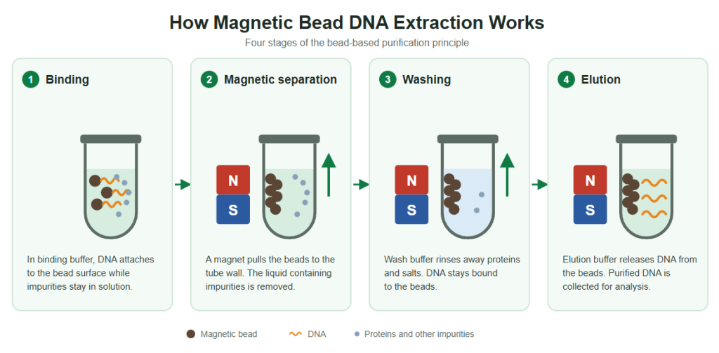
DNA binds to the beads, a magnet holds them in place, impurities are washed away, and purified DNA is eluted.
The method relies on a simple principle: under the right buffer conditions, DNA binds reversibly to the surface of magnetic beads, and a magnet holds the beads in place while everything else is removed.
Binding. After the sample is lysed, a binding buffer is added. In many silica-based systems, chaotropic salts and alcohol promote DNA binding to the bead surface. Other systems use carboxyl-coated beads with a polyethylene glycol (PEG) and salt mixture to bind DNA.
Magnetic separation. A magnet pulls the beads, with DNA attached, to the side or bottom of the tube or well. The remaining liquid, containing proteins, salts and other contaminants, is removed.
Washing. The beads are washed, usually with an ethanol-based wash buffer, to remove residual impurities while DNA stays bound.
Elution. The beads are resuspended in a low-salt elution buffer or water, which releases the DNA. The magnet then holds the beads back while the purified DNA is transferred.
The beads themselves are small superparamagnetic particles, which means they respond strongly to a magnet but do not stay magnetized once the magnet is removed. This allows them to be resuspended evenly and reused across many mixing and separation cycles within a protocol. For kit developers and laboratories that need the particles themselves, magnetic microspheres can be used as the solid phase for nucleic acid extraction and purification.
Step-by-Step Magnetic Bead DNA Extraction Workflow
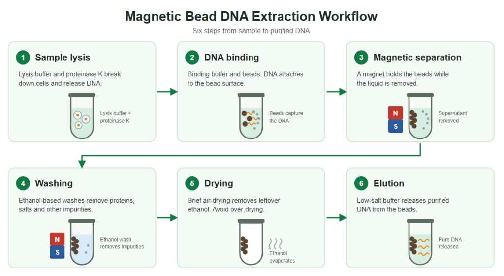
A typical workflow follows these stages. Exact volumes, times and temperatures depend on the kit or protocol being used.
The six main steps of magnetic bead DNA extraction.
Step 1: Sample lysis
A lysis buffer disrupts cell membranes and releases DNA. Depending on the sample, lysis may include detergents, chaotropic salts, mechanical disruption or enzymatic pretreatment. For tissue samples, a dedicated option is the Magnetic Fast Tissue DNA Kit.
Step 2: Protein digestion
For many sample types, proteinase K is added during lysis to digest proteins, release DNA from protein complexes and inactivate nucleases. If you want a detailed explanation of this step, see our guide to proteinase K in DNA extraction.
Step 3: DNA binding to magnetic beads
The lysate is mixed with beads and binding buffer. Thorough mixing and sufficient binding time allow DNA to attach to the bead surface. Many general-purpose kits follow this lysis, binding, washing and elution format for common sample types.
Step 4: Magnetic separation
The tube or plate is placed on a magnetic stand, and the beads collect against the wall. The supernatant is carefully removed without disturbing the bead pellet.
Step 5: Washing
Two or more wash steps remove proteins, salts and other contaminants. Ethanol-based washes are common, and complete removal of ethanol afterwards is important, because residual ethanol can inhibit downstream reactions.
Step 6: Drying
The beads are briefly air-dried to evaporate leftover ethanol. Over-drying can make DNA harder to elute, so this step should follow the protocol closely.
Step 7: Elution
The beads are resuspended in elution buffer or nuclease-free water. After a short incubation, the beads are separated again and the purified DNA is collected for PCR, qPCR, sequencing, genotyping or other downstream analysis.
Magnetic Beads vs Spin Columns
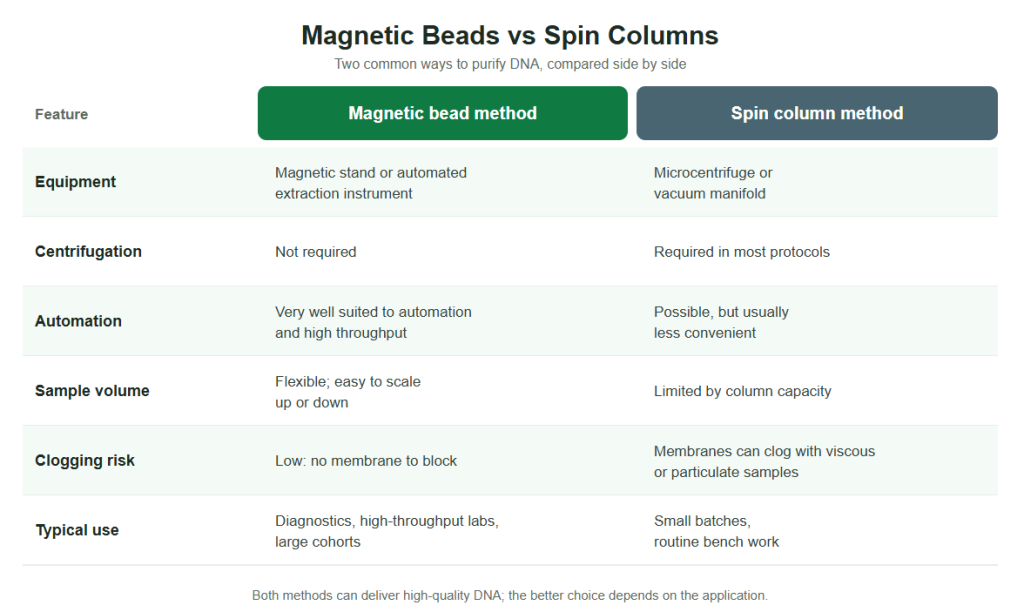
Both methods can deliver high-quality DNA, and the better choice depends on the application.
For low sample numbers and simple bench work, spin columns remain a good option. When throughput, consistency and automation matter, magnetic beads for DNA extraction is usually the more practical choice. Magnetic bead methods are also widely used for low-concentration samples such as plasma, for which the Magnetic Circulating DNA Kit is intended.
Magnetic beads suit automation and high throughput, while spin columns remain convenient for small batches.
Advantages and Limitations
Advantages
Easy to automate on extraction platforms, which reduces operator variability.
No centrifugation, so workflows are faster and easier to scale.
Flexible sample volumes and compatibility with plates and tubes.
Less risk of clogging with viscous or particulate samples.
Elution in small volumes gives concentrated DNA.
Limitations
Bead loss during supernatant removal can reduce yield if pipetting is careless.
Residual ethanol or carried-over beads can interfere with downstream reactions.
Over-drying beads can reduce elution efficiency.
Highly viscous or protein-rich samples may need extra pretreatment.
Performance depends on matching beads, buffers and the instrument.
Key Factors That Affect Yield and Purity
The following factors have the biggest effect on results. The ranges are typical values from common protocols, and the validated protocol for your kit or instrument should always take priority.
Factor
What to consider
Lysis efficiency
Incomplete lysis lowers yield. Tissue should be cut or homogenized, and tough samples may need longer or stronger digestion.
Binding conditions
Salt and alcohol concentration, pH and bead-to-sample ratio all influence how much DNA binds.
Mixing
Beads should be fully resuspended during binding so that all DNA can contact the surface.
Binding time
Several minutes is typical for many protocols. Longer times can help for dilute samples.
Magnetic separation time
Beads should be completely collected before the supernatant is removed; the required time depends on bead size and magnet strength.
Washing
Enough wash steps are needed to remove contaminants, and ethanol should be removed completely afterwards.
Drying
Beads should be dried only until residual ethanol is gone, without cracking or over-drying.
Elution
Elution volume, temperature and incubation time affect recovery. Smaller volumes give higher concentrations but may lower total yield.
Magnetic Bead DNA Extraction for Different Sample Types
Sample type
Practical considerations
Blood
Contains many proteins and, in some cases, anticoagulants. Efficient lysis and protein digestion help recover genomic DNA consistently.
Tissue
Cut or homogenize into small pieces and allow enough digestion time. A dedicated workflow such as the Magnetic Fast Tissue DNA Kit is designed around tissue samples.
Plasma and serum
Cell-free DNA is present at very low concentrations and in short fragments, so the method needs good recovery of small amounts of DNA. The Magnetic Circulating DNA Kit is intended for circulating DNA applications.
Swabs and saliva
May contain mucus and microorganisms that interfere with amplification. Proper lysis and washing are important.
Cultured cells
Usually lyse easily and give good yields with short protocols.
Bacteria and fungi
Cell walls may need extra pretreatment, such as enzymatic or mechanical disruption, before the standard workflow.
Magnetic Bead DNA Extraction in Automated and High-Throughput Workflows
Automation is the main reason magnetic bead DNA extraction dominates in diagnostics and large-scale testing. Instruments move the beads between wells of a plate, mix, separate and wash with minimal operator intervention. This improves reproducibility and shortens hands-on time, which is critical for clinical laboratories, screening programs and large cohort studies.
For automated workflows, the following points matter:
Compatibility of the beads and buffers with the instrument and plate format.
Speed, including bead collection time and the lysis step.
Reproducibility across wells, runs and operators.
Contamination control, because many samples are processed in parallel.
Reagent stability, so that kits perform consistently throughout their shelf life.
Troubleshooting Magnetic Bead DNA Extraction
Problem
Possible cause
What to try
Low DNA yield
Incomplete lysis; insufficient binding time; bead loss during supernatant removal
Make sure protein digestion is complete and wash steps are performed properly
Low A260/230 ratio
Residual salts or ethanol
Add or extend wash steps and ensure ethanol is fully removed
PCR or qPCR inhibition
Residual ethanol, salts or carried-over beads
Dry briefly, use sufficient washes, and separate beads fully before collecting DNA
Beads clump or are hard to resuspend
Over-drying; insufficient mixing
Shorten the drying step and resuspend thoroughly
Beads in the final eluate
Incomplete magnetic separation
Allow longer separation time and take eluate carefully
Degraded DNA
Nuclease activity; delayed lysis; harsh handling
Process or lyse samples promptly and avoid unnecessary shearing
Variable results between runs
Inconsistent reagents or pipetting
Check bead and reagent lot consistency and automate liquid handling where possible
How to Choose Magnetic Beads and Kits for DNA Extraction
For laboratories choosing a kit, the best option is the one validated for the sample type and throughput you need. For diagnostic reagent developers and kit manufacturers, the selection of the beads themselves is a critical design choice. Key criteria include:
Binding capacity and recovery. The beads should capture DNA efficiently across the range of input amounts in your application.
Particle size and uniformity. Consistent size gives predictable magnetic response and binding behavior.
Magnetic response. Beads should separate quickly and completely, but redisperse easily after the magnet is removed.
Dispersion and stability. Beads should resist aggregation and remain stable during storage.
Surface chemistry. The surface must match the binding chemistry of your buffer system, for example silica-based or carboxyl-based.
Purity and contamination control. Residual nucleases or impurities can affect sample integrity and downstream assays.
Lot-to-lot consistency. A protocol validated with one lot should perform in the same way with later lots.
Scale-up and supply. Suppliers should be able to support the move from development to bulk manufacturing, with reliable supply and quality documentation.
Hyasen Magnetic Bead Products for DNA Extraction
Hyasen offers magnetic bead products for different needs, from raw materials for kit developers to ready-to-use extraction kits:
Magnetic Microspheres: magnetic beads that can be used as the solid phase for nucleic acid extraction and purification.
Please refer to each product page for specifications, applicable sample types and instrument compatibility.
Frequently Asked Questions
What is magnetic bead DNA extraction?
Magnetic bead DNA extraction is a method in which DNA binds to magnetic particles, the particles are held with a magnet while impurities are washed away, and purified DNA is then eluted. It does not require centrifugation, which makes it easy to automate.
How does magnetic beads for DNA extraction differ from spin column extraction?
Spin columns capture DNA on a membrane and rely on centrifugation or vacuum to move liquids through it. Magnetic beads capture DNA in solution and use a magnet to separate it, which is better for automation and flexible sample volumes.
Is magnetic bead DNA extraction better than column-based methods?
It depends on the application. Magnetic beads are usually preferred for high-throughput and automated work, while spin columns remain convenient for small batches at the bench.
Do you need proteinase K for magnetic beads for DNA extraction?
Not always, but it is commonly used in the lysis step for protein-rich samples such as blood and tissue, because it digests proteins and reduces nuclease activity.
What can cause low DNA yield with magnetic beads?
Common causes include incomplete lysis, insufficient binding time or mixing, bead loss during supernatant removal, and over-drying of the beads before elution.
Why is there ethanol or bead carryover in my eluate?
Residual ethanol comes from incomplete removal of the wash buffer, and bead carryover comes from incomplete magnetic separation before collecting the eluate. Allow enough drying and separation time.
Can magnetic bead extraction be used for circulating DNA?
Yes. Magnetic bead methods are widely used for cell-free and circulating DNA, provided the protocol is designed to recover low concentrations of short fragments efficiently.
Can the same beads be used for DNA and RNA?
Many bead chemistries can bind both DNA and RNA, but the buffers and steps differ, so the protocol should be designed or validated for the target nucleic acid.
What should manufacturers consider when sourcing magnetic beads?
Binding capacity, particle uniformity, magnetic response, dispersion, surface chemistry, purity, lot-to-lot consistency and supply capacity are the key factors.
Conclusion
Magnetic bead DNA extraction offers a fast, scalable and automation-friendly way to obtain high-quality DNA from a wide range of samples. Good results depend on efficient lysis, correct binding and washing conditions, careful magnetic separation and the right choice of beads and reagents for each application.
Looking for magnetic beads or DNA extraction kits for your laboratory or product development? Contact Hyasen to discuss samples, technical specifications, bulk supply and customized solutions.
PROWESS®LAMINOPLASTY SYSTEM
The PROWESS System provides a comprehensive solution for Laminoplasty procedures and consists of streamlined instrumentation and a variety of plate and spacer options for intraoperative adaptability. The instrumentation are intuitive and maintain secure connections to the implants. The implants are offered in a range of sizes and are color coded for easy visual identification.
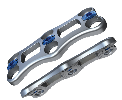
Prowess Spacer Plates
4-12mm lengths (in 2mm increments) that work with various Spacers
8mm across neck, 5.7mm across center graft window
Color-coded plates allow for easy identification
Double-bend design, low-profile to allow further contouring
Anterior ridges resist slippage and increase torsional stability
Accepts Ø2.3 x 4mm Self-Tapping Bone Screw for assembly
Prowess Hook Plates
4-12mm lengths (in 2mm increments) that work with various Spacers
8mm across neck
Color-coded plates allow for easy identification
Double-bend design, low-profile to allow further contouring
Anterior ridges resist slippage and increase torsional stability
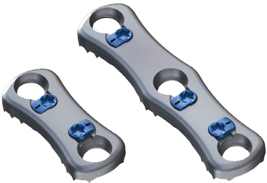
Prowess Hinge Plates
8mm across neck
Singe-bend design, low-profile to allow further contouring
Anterior ridges resist slippage and increase torsional stability
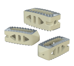
Prowess Spacers
Titanium or PEEK construction
6mm wide, and 4mm minimum height from posterior face to anterior curve
Implant Inserter grip features
Medial, lateral, and anterior anatomical curvatures
Graft cavities on all sizes
Accepts Ø2.3 x 4mm Self-Tapping Bone Screw for assembly
Titanium Spacers are color-coded for easy identification
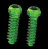
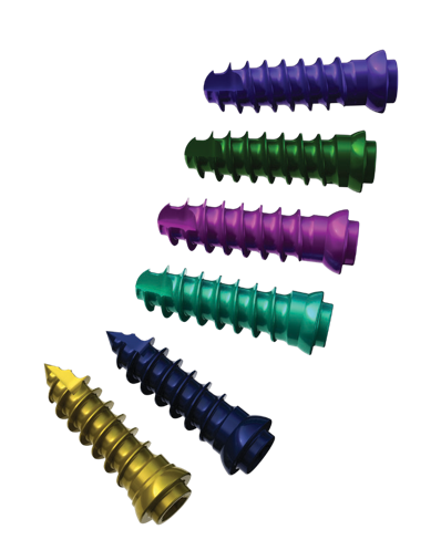
Prowess Screws
Ø2.3 and Ø2.7 in 4-12mm lengths (in 2mm increments)
Self-Drilling and Self-Tapping
Color-coded screws allow for easy identification
Works with self-retaining Screw Driver
Same Ø2.3 x 4mm Self-Tapping bone screw assembles Spacer to Spacer Plate
CuriGraft Viable Allogenic Bone Matrix
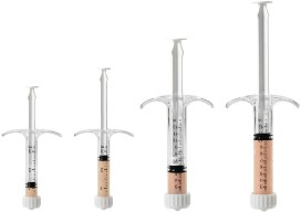
CuriGraft® Viable Allogenic Bone Matrix is the next generation allograft containing the three key elements ideal for bone formation, offering a variety of clinical applications, including the spine, upper extremity, foot and ankle and orthopedic oncology.
Key Features
Osteoconductive three-dimensional scaffold with cortical and cancellous components
Demineralized cortical bone scaffold. Demineralized cortical bone has been identified to have osteoinductive potential1
Viable endogenous bone cells to support osteogenic healing processes
DMSO-FREE cryoprotectant, requiring no rinsing or decanting
Proprietary mixture of mineralized cancellous bone along with demineralized cortical fibers
Easy-to-use syringe, just thaw and use!
Four-hour working window for implantation post thaw
Storage: -65°C or colder
Size
Product Code
1.0cc
C034-0001-MCBMV
2.5cc
C034-0025- MCBMV
5.0cc
C034-0005- MCBMV
10.0cc
C034-0010- MCBMV
Reference: 1) Gruskin, E. et.al., Demineralized bone matrix in bone repair: history and use. Advanced Drug Delivery Reviews, 2012. 64:1063-1077 Biologics
CuriGraft® DBM Putty - Demineralized Bone Matrix Putty
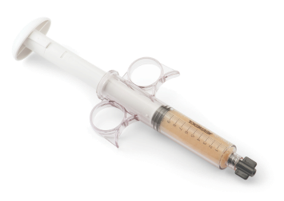
CuriGraft® DBM Putty is a demineralized bone putty matrix indicated for use in the extremities, posterolateral spine, and pelvis as a bone void filler or for treatment of osseous defects.
Key Features
Product is provided in a ready-to-use syringe
Equivalent to iliac crest autograft proven in a preclinical spinal fusion model1
Assured osteoinductivity via validated processing
Cohesive handling that resists irrigation due to robust biocompatible carrier
Patient safety ensured through terminal sterilization and viral inactivation
Cortical demineralized bone and a carboxymethyl cellulose (CMC) carrier.
Size
Product Code
1.0cc
C024-0001-PTYV
2.5cc
C024-0025-PTYV
5.0cc
C024-0005-PTYV
10.0cc
C024-0010-PTYV
Reference: 1) Kiely, P.D. et al., (2014) Evaluation of a new formulation of demineralized bone matrix putty in a rabbit posterolateral spinal fusion model. The Spine Journal. September 14(9): 2155-2163.
SI-LUTION
The SI-LUTION® Sacroiliac Joint Fusion System is designed to offer immediate stability and long-term fixation across the sacroiliac (SI) joint. The implants are machined out of Titanium and incorporate the proprietary TRU-LOK® micro-textured surface technology. The cannulated system offers screws in multiple diameters and various lengths with optional static or kinetic stabilization caps to provide an ideal solution for every patient.
Key Features
Manufactured of Titanium with proprietary surface texturing
Large centralized helical fenestrations allow for direct allograft integration
Self-harvesting bone channels to harvest and distribute local bone during insertion
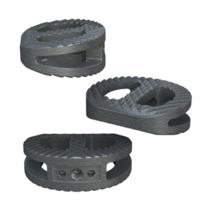
The Savant Anterior Lumbar (ALIF) Interbody Fusion System features three footprints in various heights and lordotic angles offering an ideal fit for every patient.
System Features
Manufactured of Titanium Featuring TRU-LOK®
Open Architecture
Micro-roughness surface texturing produces superior implant contact
Comprehensive discectomy instruments
Three Footprints (mm): 30W x 24D, 35W x 26D, 40W x 28D
8 – 20mm, offered in 2mm increments
Lordosis: 6° and 12°
Prodigy MIS
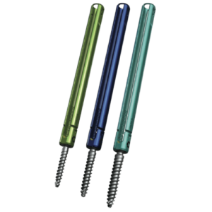
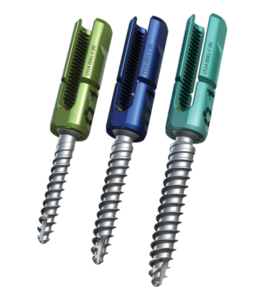
The Prodigy MIS Pedicle Screw System is designed to minimize the complexity of procedural steps while correcting and stabilizing the posterior thoracolumbar spine through a percutaneous approach. This system provides the same strength and rigidity as the Prodigy Polyaxial Screw System.
PEDICLE SCREW SYSTEM
Key Features
Minimized soft tissue disruption
Strong interface between rod and rod inserter
Undercut square thread designed to prevent cross-threading and tulip head splaying
Simple and efficient instruments provide advanced manipulation techniques
Available in 4.5, 5.5, 6.5 and 7.5 Diameter
Heights: 30-55mm in 5mm increments
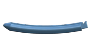
MIS ROD
Key Features
Bulleted tip to facilitate placement
Robust instrument engagement feature
PRODIGY™
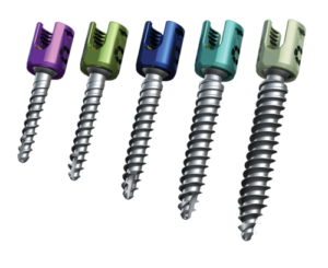
The Prodigy Polyaxial Pedicle Screw System is a posterior spinal fixation system designed to enhance the surgical experience by providing surgeons with its comprehensive implant offering and ergonomically designed instrumentation. The low-profile, friction fit design is offered in both standard and reduction geometries.
PRODIGY PEDICLE SCREW SYSTEM
Key Features
Self-tapping double lead bone screw threads
Continuous friction to maintain tulip position
Optimized instrument ports
Streamlined rod capture
Square thread designed to prevent cross-threading
Available in standard and dual-thread cortico-cancellous options
4.5 – 10.5mm diameter
Height: 25mm – 110mm lengths
5.5mm pre-bent or straight rod available
PRODIGY REDUCTION PEDICLE SCREWS
Key Features
Self-tapping double lead bone screw threads
Continuous friction to maintain tulip position
Optimized instrument ports
Streamlined rod capture
Square thread designed to prevent cross-threading
Extended tulip height of 23.5mm
26mm of threads
Reduction screws offered in 5.5 – 7.5mm in 25mm – 55mm lengths
5.5mm pre-bent or straight rod available
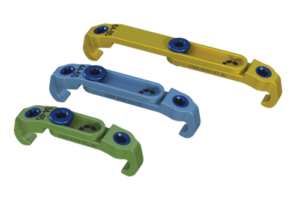
VARIABLE CROSSLINKS
Key Features
Versatile crosslink design with adjustable lengths and angle variability
Slim profile design
Center and lateral set screw features the same T20 hexalobe drive
64-85mm (Gold)
44-65mm (Light Blue)
34-45mm (Green)
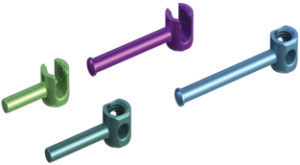
OFFSET CONNECTORS
Key Features
20, 25 and 30 open and closed Offset Connectors
Connects to a Ø5.5mm rod
Accepts the same set screw as the pedicle screws
Flanged and smooth options
Savant TLIF
The Savant Transforaminal Lumbar (TLIF) Interbody Fusion System is offered in three different material types, multiple footprints, heights, and lordosis providing multiple options for the surgeon. The convex design offers a more accurate fit to the patient’s anatomy and offers oversized, central lateral graft windows for biologic seeding. The Savant Interbody Fusion System offers Comprehensive discectomy instruments and features instrumentation that allows for better control and more precise implant placement.
SAVANT PEEK TLIF
Key Features
Manufactured of biocompatible PEEK polymer
Strategically positioned tantalum radiopaque markers to optimize visibility and placement
Angled lateral windows on anterior side of implant to retain bone graft material
Large back surface area for increased instrument interface
Footprints (mm): 28L x 9W, 28L x 11W, 32L x 9W and 32L x 11W
Heights: 6 – 16mm, offered in 1mm increments
Lordosis: 0° or 8º
SAVANT TITANIUM TLIF
Key Features
Manufactured of Titanium Featuring TRU-LOK®
Open Architecture
Designed to offer immediate stability and long-term fixation
Micro-roughness surface texturing produces superior implant contact
Footprints (mm): 28L x 9W, 28L x 11W, 32L x 9W and 32L x 11W
Heights: 6 – 16mm, offered in 1mm increments
Lordosis: 0° or 8°
SAVANT TITANIUM PLASMA COATED TLIF
Key Features
Manufactured of PEEK with a titanium plasma spray coating
Footprints (mm): 28L x 9W, 28L x 11W, 32L x 9W and 32L x 11W
Heights: 6 – 16mm, offered in 1mm increments
Lordosis: 0° or 8º
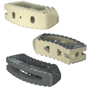
Savant PLIF
The Savant Posterior Lumbar (PLIF) Interbody Fusion System self-distracting, bulleted tip is offered in three different material types, multiple footprints, heights, and lordosis providing multiple options for the surgeon. The Savant PLIF Interbody Fusion System features instruments that are simple and intuitive to use.
SAVANT PEEK PLIF
Key Features
Manufactured of biocompatible PEEK polymer
Strategically positioned tantalum radiopaque markers to optimize visibility and placement
Multi-directional teeth resist migration
Footprints (mm): 22L x 10W, 26L x 10W and 30L x 10W
Heights: 6 – 16mm, offered in 1mm increments
Lordosis: 0° and 8
SAVANT TITANIUM PLIF
Key Features
Manufactured of Titanium Featuring TRU-LOK®
Open Architecture
Micro-roughness surface texturing produces superior implant contact
Footprints (mm): 22L x 10W, 26L x 10W and 30L x 10W
Heights: 6 – 16mm, offered in 1mm increments
Lordosis: 0° or 8°
SAVANT TITANIUM PLASMA COATED PLIF
Key Features
Manufactured of PEEK with a titanium plasma spray coating
Optimal Radiolucency
Increased surface topography
Footprints (mm): 22L x 10W, 26L x 10W and 30L x 10W
Heights: 6 – 16mm, offered in 1mm increments
Lordosis: 0° and 8°
Aptitude
The Aptitude Cervical Plates feature a slim profile design allowing for a smooth anterior service. The simple and intuitive one-step locking mechanism facilitates complete control by the surgeon during the procedure. The Aptitude system offers streamlined instrumentation to accommodate the needs of the patients and surgeons.
ANTERIOR CERVICAL PLATES
Key Features
Low-profile Plate
Large Graft Window
Simple and effective, one-step locking mechanism
Pre-Lordosed to match patient anatomy
1-5 level plates
17mm plate width
2.2mm thick
ANTERIOR MIDLINE CERVICAL PLATES
Key Features
Small incision, minimizing lateral retraction
Narrow width optimizes post-operative visualization of the fusion site
Posterior cleats resist slippage and increase torsional stability
1-2 level plates
10.5mm plate width
2.2mm thick
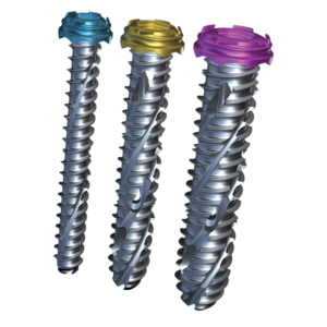
APTITUDE SCREWS
Key Features
Ø4.0 and Ø4.5 in 12-18mm lengths (in 2mm increments)
Self-Drilling, Self-Tapping, Fixed and Variable Screws
Color-coded screws allow for easy identification
Same screws used for both Standard and Midline plates
Sage
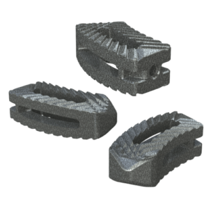
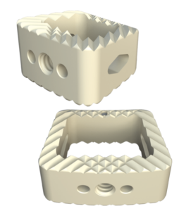
The Sage Cervical Interbody Fusion System is designed to support cervical loads while maximizing surface between the implant and vertebral bodies. The Interbody is offered in four footprints to accommodate individual patient anatomy while the streamlined instrumentation is designed to aid in consistent and accurate implant placement.
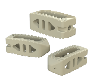
SAGE PEEK CERVICAL
Key Features
Manufactured of biocompatible PEEK polymer
Tantalum radiopaque markers to optimize visibility and placement
Multi-directional aggressive teeth designed to prevent implant migration
Large, central graft cavity for packing bone graft
Color-coded instruments for easy identification
Footprints (mm): 12W x 11D, 14W x 12D, 16W x 13D, and 18W x 14D
Heights: 5-12mm, offered in 1mm increments
Lordosis: 7°
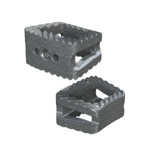
SAGE TITANIUM CERVICAL
Key Features
Manufactured of Titanium with surface texturing
Open Architecture
Micro-roughness surface texturing produces superior implant contact
Designed to ensure a high degree of compressive strength and dimensional stability
Footprints (mm): 12W x 11D, 14W x 12D, 16W x 13D, and 18W x 14D
Heights: 5-12mm, offered in 1mm increments
Lordosis: 7°
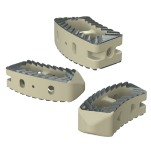
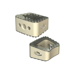
SAGE TITANIUM COATED CERVICAL
Key Features
Manufactured of PEEK with a titanium plasma spray coating
Surface topography produces superior implant contact and construct stability
Optimal Radiolucency
Self-distracting leading edge to facilitate implant insertion
Footprints (mm): 12W x 11D, 14W x 12D, 16W x 13D, and 18W x 14D
Heights: 5-12mm, offered in 1mm increments
Lordosis: 7°
Contact Curiteva
Contact us to learn more about Curiteva’s products and technology





 System Features
 Key Features
 Key Features
 Key Features
 Key Features
 Key Features
 Key Features
 Key Features
 Key Features
 Key Features