How to Choose the Right Interbody Fusion Implant?

Choosing the right interbody fusion implant begins with the patient, not the product catalogue.

Lumbar fusion procedures have increased substantially in the United States. A review of national inpatient data reported a 67.3% rise between 2001 and 2011 (Rajaee et al., Spine, 2012). That growth makes implant selection more important, not automatically easier. A device must match anatomy, pathology, bone quality, surgical access, and the intended fusion strategy.

“The implant is only one part of the fusion procedure,” says spine surgeon Dr. Juan S. Uribe, a recognized specialist in complex spinal reconstruction. His point deserves attention. Cage geometry cannot correct poor endplate preparation, unstable fixation, or an unsuitable surgical indication.

The U.S. Food and Drug Administration emphasizes that spinal implants require careful evaluation of risks, benefits, materials, and patient-specific factors (FDA, Guidance for Industry: Spinal System Guidance, 2021). The North American Spine Society also stresses evidence-based decision-making when assessing lumbar fusion technologies (NASS Clinical Guidelines).

Look closely at the details. A narrow cage may simplify insertion, yet provide limited surface contact. A larger footprint may improve load distribution, but increase preparation demands. Subsidence remains a practical concern, especially when bone density is poor.

There is no perfect implant.

Even experienced surgeons can reassess a familiar choice after reviewing imaging, alignment, and bone health. This article examines implant materials, footprint, height, lordotic angle, insertion technique, and clinical evidence. The goal is not to promote one design. It is to support a safer, more transparent interbody fusion decision.

How to Choose the Right Interbody Fusion Implant?

Define the Diagnosis and Goals of Interbody Fusion

How to Choose the Right Interbody Fusion Implant?

Define the Diagnosis and Goals of Interbody Fusion

Implant selection should begin with a precise diagnosis, not a preferred device. Confirm whether pain comes from instability, disc degeneration, deformity, stenosis, or nonunion. The Global Burden of Disease study estimated 619 million people experienced low back pain in 2020. That figure describes a major burden, not automatic surgical need. Imaging must match symptoms, neurological findings, and functional limitations.

The surgical goal should be equally specific. Fusion may aim to restore disc height, decompress a nerve, correct alignment, or stabilize painful motion. Each goal changes the preferred implant shape, footprint, height, and material. NASS evidence-based recommendations emphasize correlating radiographic findings with clinical symptoms. A cage that looks ideal on a scan may still fail to address the patient’s main complaint. This is where planning can become imperfect. Surgeons should question whether fusion offers more value than decompression, rehabilitation, or continued observation.

Tips: Write the diagnosis in one sentence. Then write the surgical goal in another. Review standing radiographs, MRI, and bone quality before choosing dimensions. Consider osteoporosis, smoking, diabetes, prior surgery, and sagittal alignment. The FDA’s post-market surveillance resources also show why implant safety requires ongoing monitoring. A sound plan includes expected benefits, possible complications, and a measurable follow-up target. Sometimes, the best decision is to delay implantation.

Compare Implant Designs, Materials, and Surgical Approaches

How to Choose the Right Interbody Fusion Implant?

Compare Implant Designs, Materials, and Surgical Approaches

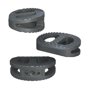
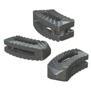
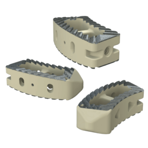
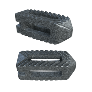
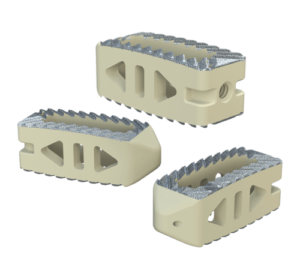
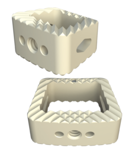
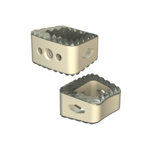
Choosing an interbody implant starts with the patient’s anatomy, not the implant catalogue. A wider footprint may improve stability and reduce subsidence risk. However, it can require more extensive preparation of the vertebral endplates. Lordotic implants may help restore alignment, especially in carefully selected lumbar cases. Their value depends on placement, bone quality, and overall spinal balance.

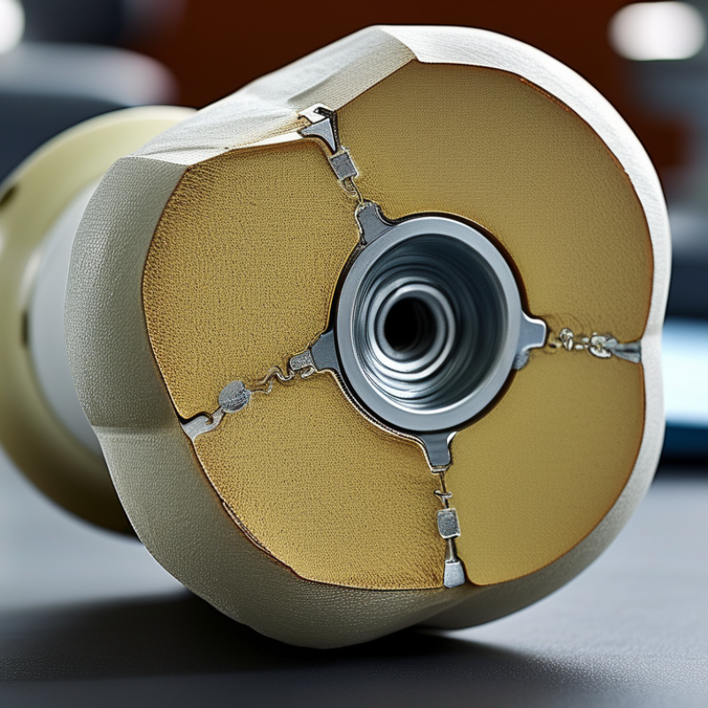
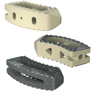
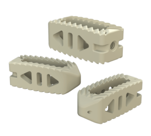
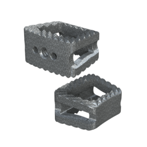
Material selection also involves trade-offs. PEEK implants can provide radiolucent imaging and allow clearer assessment of fusion. Titanium implants may offer strong fixation and surface features that support bone attachment. Porous structures can encourage bone growth, but they do not eliminate nonunion risk. No material is ideal. Imaging needs, osteopenia, and revision planning should influence the decision.

The surgical approach changes both access and risk. Anterior approaches may improve disc height while introducing vascular or abdominal considerations. Lateral approaches can reduce posterior muscle disruption, yet the psoas and nearby nerves require careful evaluation. Posterior approaches provide familiar access but may involve more muscle handling. Patient positioning, prior surgery, and nerve anatomy matter greatly. A practical review should include CT findings, MRI details, bone density, alignment goals, and the surgeon’s documented experience. The most attractive design on paper may perform poorly in the wrong anatomy. That uncomfortable possibility deserves honest discussion.

Match Implant Size and Shape to the Patient’s Anatomy

How to Choose the Right Interbody Fusion Implant?

Match Implant Size and Shape to the Patient’s Anatomy

Choosing an interbody fusion implant begins with understanding the patient’s three-dimensional anatomy. Plain radiographs show alignment, but CT scans reveal endplate shape, disc height, and bony defects. MRI may clarify nerve compression and disc degeneration. These details guide implant selection more reliably than measurements alone.

A properly sized implant should contact strong portions of the endplates without breaching the cortical margins. It must restore disc height without creating excessive tension. Width matters too. A narrow implant may provide limited support, while an oversized implant can damage the endplate. Small differences matter. The surgeon should also consider segmental lordosis, pelvic parameters, and the patient’s natural spinal curve. Shape should follow the anatomy, not force it into a predetermined form.

Bone quality can change the plan. Osteoporotic bone may require careful preparation and gentler insertion. During trialing, resistance and seating depth provide valuable feedback. Yet intraoperative findings can challenge the original imaging-based plan. No sizing plan is flawless. A measurement can mislead. Rechecking implant position with imaging may prevent avoidable errors. I have found that conservative preparation often protects the endplate better than aggressive distraction. Still, every case has uncertainty, especially when anatomy is asymmetric or severely collapsed. The final choice should reflect imaging, surgical experience, patient-specific risks, and the implant’s intended biomechanical role.

Evaluate Clinical Evidence, Risks, and Patient-Specific Factors

Choosing an interbody fusion implant requires more than comparing shape, material, or marketing claims. Clinical evidence should guide the decision. Look for peer-reviewed studies with meaningful follow-up periods, not only early fusion rates. Review outcomes such as pain improvement, neurological recovery, subsidence, nonunion, revision surgery, and patient-reported function. Evidence is rarely perfect. Some studies use small groups or different surgical techniques, making direct comparisons difficult.

Risks must be considered alongside expected benefits. Possible complications include infection, nerve injury, implant migration, subsidence, nonunion, and adjacent-segment problems. Patient-specific factors can change those risks. Low bone density may increase subsidence risk. Smoking and poorly controlled diabetes may impair healing. Spinal alignment, bone quality, anatomy, previous surgery, and daily activity also matter. A technically successful operation may still disappoint if expectations are unrealistic.

Tips: Ask the surgeon why a specific implant fits your anatomy. Request evidence for similar patients. Discuss bone-density testing when appropriate. Ask how complications are managed. Bring a complete medication and medical history. The cheapest option is not always the safest, and the newest option is not automatically better. A careful decision often depends on details that imaging alone misses. Pain patterns, work demands, and recovery support deserve attention too.

Confirm Compatibility, Surgical Planning, and Follow-Up Needs

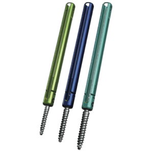
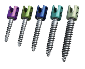
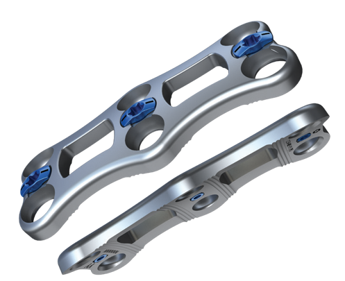
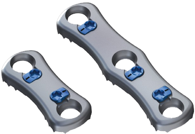
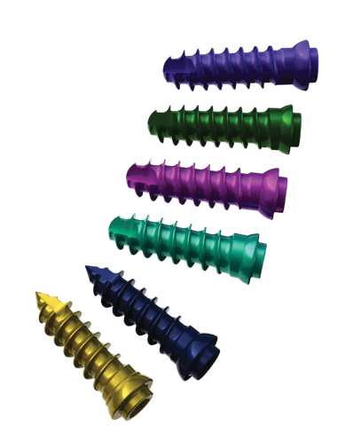
Choosing an interbody fusion implant requires more than comparing sizes. Compatibility starts with the patient’s anatomy, diagnosis, bone quality, and planned surgical approach. The implant should match the prepared disc space and work with the fixation system selected for that level. Surgeons should review CT or MRI findings, standing radiographs, and relevant medical history before making a final choice. Material sensitivities, previous operations, and imaging requirements also deserve attention. Small mismatches can complicate placement. That risk is easy to underestimate.

Surgical planning turns compatibility into a practical sequence. The team should confirm implant dimensions, insertion tools, trial sizes, positioning targets, and backup options before incision. A detailed plan can reduce avoidable delays, but it cannot predict every finding. Scar tissue, poor visualization, or weaker bone may require an adjustment. No checklist catches everything. The surgeon should explain these possibilities, expected benefits, limitations, and alternatives in understandable language. Nurses and technicians also need the same plan. Communication gaps matter.

Follow-up needs should influence implant selection before surgery, not afterward. Patients may need scheduled examinations and radiographs to assess alignment, subsidence, hardware position, and signs of fusion. The schedule depends on the procedure, symptoms, and clinical judgment. New weakness, fever, wound drainage, or worsening pain requires prompt medical contact. A follow-up plan should identify who reviews images and how concerns are handled. This is often overlooked. Reliable records help compare healing over time. If progress is uncertain, the care team may reconsider activity limits, additional imaging, or further evaluation.