| Standard RGB Imaging | Visible-light camera with computer-vision and machine-learning analysis | Approximately 1–5 seconds | Visible spots, redness, pore appearance, wrinkles, skin tone distribution, facial symmetry, and texture patterns | Affordable, fast, non-contact, easy to integrate with mobile or clinic workflows, and widely supported by image-analysis software | Results can change with lighting, camera distance, makeup, skin moisture, and image quality; it does not directly measure deeper tissue properties | Routine skin consultations, progress tracking, education, and high-volume retail or clinical screening | High |
| Cross-Polarized Imaging | Visible-light camera combined with cross-polarizing filters to reduce surface glare | Approximately 2–8 seconds | Redness, vascular appearance, pigmentation distribution, uneven tone, and low-contrast surface features | Improves image consistency by reducing specular reflection and can reveal color differences that are difficult to see under ordinary lighting | Still affected by exposure settings and skin preparation; primarily evaluates optical appearance rather than biological activity | Detailed pigmentation and redness assessment, treatment documentation, and before-and-after comparison | High |
| Multispectral Imaging | Several controlled visible and near-infrared wavelength bands captured by calibrated sensors | Approximately 5–20 seconds | Relative pigmentation, erythema, melanin-related patterns, surface condition, and selected subsurface optical indicators | Provides more objective spectral information than ordinary RGB imaging and supports standardized longitudinal measurements | Higher equipment cost, calibration requirements, and results that depend on validated algorithms and controlled acquisition conditions | Professional skin assessment, research-supported cosmetic evaluation, and treatment monitoring | Very High |
| 3D Structured-Light or Depth Imaging | Projected light pattern or depth sensor used to reconstruct facial geometry | Approximately 1–10 seconds | Facial contours, volume, symmetry, surface relief, wrinkle depth, folds, and selected dimensional changes | Captures shape and topography that cannot be reliably measured from a flat photograph; useful for objective geometry tracking | Movement, facial expression, hair, reflective surfaces, and head-position changes can reduce repeatability | Wrinkle-depth assessment, facial contour analysis, aesthetic treatment planning, and volumetric progress tracking | Very High |
| Near-Infrared Imaging | Near-infrared illumination and sensors that detect reflected or partially penetrating light | Approximately 3–15 seconds | Optical differences associated with pigmentation, vascular patterns, hydration-related contrast, and selected subsurface features | Can provide information that is less dependent on visible-light appearance and may improve detection of low-contrast patterns | Interpretation is indirect; measurements require careful calibration and should not be treated as a medical diagnosis without clinical validation | Advanced cosmetic consultation, research, and complementary analysis alongside visible-light imaging | High |
| Fluorescence Imaging | Excitation light and camera detection of emitted fluorescence from the skin surface | Approximately 2–10 seconds | Surface fluorescence patterns, selected porphyrin-related signals, oil-associated areas, and surface irregularities | Highlights features that may be difficult to distinguish under ordinary visible illumination | Signals can be influenced by cleansing, cosmetics, ambient light, and individual skin characteristics; interpretation requires trained users | Surface-condition education, oil-area visualization, and supplementary professional assessment | Specialized |
| Thermal Imaging | Infrared thermal sensor measuring emitted surface temperature patterns | Approximately 5–30 seconds | Relative temperature distribution, thermal asymmetry, and changes associated with surface blood-flow patterns | Contact-free and capable of showing dynamic thermal changes that are not visible in ordinary photographs | Strongly affected by room temperature, airflow, recent exercise, hot or cold drinks, and acclimatization time | Specialized research, complementary inflammation-related observation, and controlled clinical studies | Specialized |
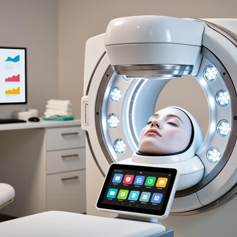
| Hybrid Multimodal Systems | Combination of RGB, polarized, multispectral, depth, or other calibrated sensors in one workflow | Approximately 5–30 seconds | Combined appearance, color, texture, geometry, and selected optical indicators with longitudinal scoring | Offers the broadest information set and can cross-check findings across different sensing methods | Higher purchase and maintenance cost, greater workflow complexity, data-standardization challenges, and a stronger need for user training | Comprehensive professional assessment, clinics with standardized protocols, and high-value treatment planning | Best Overall Potential |
| AI-Assisted Analysis Layer | Machine-learning models applied to images and measurements from one or more sensors | Usually less than 10 seconds after image capture | Feature segmentation, severity scoring, change detection, personalized recommendations, and risk flags for human review | Improves consistency, automates reporting, supports trend analysis, and can combine multiple measurements into an easier-to-read result | Accuracy depends on training data, demographic coverage, image quality, validation methods, privacy controls, and human oversight | Standardized reporting, personalized skincare planning, and longitudinal client monitoring | Core Development Area |







 System Features
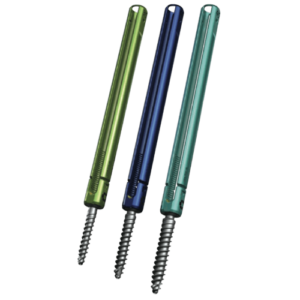 Key Features
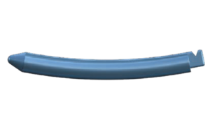 Key Features
 Key Features
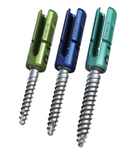 Key Features
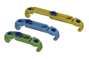 Key Features
 Key Features
 Key Features
 Key Features
 Key Features